Intraoperative Cancer Visualization 2026: Real-Time Surgical Oncology and Tumor Margin Detection
Intraoperative cancer visualization is transforming surgical oncology by enabling real-time detection of tumor margins using fluorescence imaging, molecular dyes, and AI-based optical systems. This technology reduces re-operation rates and improves surgical accuracy, though challenges remain in clinical interpretation and workflow integration.
The Glowing Margin: Shifting Surgical Oncology from Blind Touch to Real-Time Sight
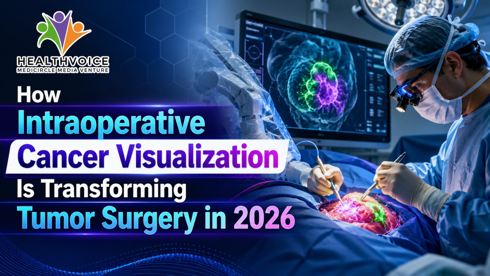
How Intraoperative Cancer Visualization Is Transforming Tumor Surgery in 2026
For nearly a century, surgical oncology has relied on visual inspection, tactile feedback, and delayed histopathology reports to determine whether a tumor has been fully removed. Surgeons would excise suspected cancer tissue, send it for analysis, and wait days to confirm whether the margins were clean.
In 2026, this retrospective model is being replaced by a new paradigm: real-time intraoperative cancer visualization. Using fluorescence imaging, molecular tracers, and AI-powered optical systems, surgeons can now “see” cancer at the microscopic level during surgery itself.
This shift is dramatically reducing re-operation rates, improving surgical precision, and redefining the future of cancer surgery.
The Limitations of Traditional Surgical Oncology
Why Cancer Surgery Relied on Delayed Pathology
In conventional tumor resection, surgeons depend on:
* Visual identification of abnormal tissue* Manual palpation of tumor boundaries* Post-surgical histopathology (5–7 day delay)
The Margin Problem
A “negative margin” indicates no cancer cells at the edge of the removed tissue, while a “positive margin” means residual cancer remains.
This delay creates critical issues:
* Uncertainty during surgery* Higher risk of repeat operations* Delayed treatment adjustments* Increased patient anxiety
The Rise of Real-Time Intraoperative Cancer Visualization
From Guesswork to Live Tumor Detection
Modern surgical oncology is transitioning into an era of real-time molecular imaging, where cancer is visualized during the operation itself.
Instead of relying on post-operative analysis, surgeons now use advanced imaging systems to detect tumor boundaries instantly.
Key Technologies Powering Intraoperative Cancer Imaging
1. Fluorescence Lifetime Imaging (FLIm)
FLIm enables label-free tumor detection by analyzing how tissues emit and decay light after laser excitation.
How it works:
* Low-energy laser pulses are applied to tissue* Measures nanosecond fluorescence decay rates* Detects metabolic differences between healthy and cancerous cells
Clinical advantage:
* Real-time 20 fps imaging* Color-coded tumor probability maps* High sensitivity to metabolic abnormalities
2. Targeted Molecular Fluorescent Dyes
Making Cancer Cells Glow During Surgery
This technique uses tumor-specific dyes that bind to cancer biomarkers.
Example applications:
* PSMA-targeted dyes in prostate cancer* Near-infrared fluorescent imaging in urological oncology
Clinical advantage:
* Microscopic metastases become visible* Tumors glow under specialized imaging systems* Improved detection of hidden cancer spread
3. AI-Enabled Optical Coherence Tomography (OCT)
Deep Tissue Imaging With Artificial Intelligence
AI-powered OCT systems provide high-resolution cross-sectional imaging of tissue structures.
Key features:
* Imaging depth up to 2mm* Deep learning-based tissue classification* Fast intraoperative margin analysis (minutes, not days)
Clinical advantage:
* Real-time decision support in the operating room* Reduced dependence on delayed pathology reports
4. Specimen PET-CT Imaging Systems
Metabolic Mapping of Excised Tumors
Mobile PET-CT systems allow surgeons to scan removed tissue immediately after excision.
Technology highlights:
* Uses radiotracers such as 18F-FDG* Provides 3D metabolic imaging of tumors* Identifies residual malignant tissue in specimens
Clinical advantage:
* Detects hidden positive margins before closure* Enhances intraoperative surgical accuracy
Clinical Impact on Surgical Oncology Outcomes
Reducing Positive Margins and Re-Operations
Positive margin rates remain a major challenge in cancer surgery, especially in:
* Breast cancer (lumpectomy procedures)* Prostate cancer (radical prostatectomy)
Historical challenges:
* 21%–30% positive margin rates in breast surgery* ~23% recurrence risk in prostate cancer due to residual disease
How Real-Time Imaging Changes Outcomes
Recent clinical data shows significant improvements:
* Breast cancer surgical success rates increased from 83.3% to 95.2%* Overall diagnostic accuracy improved up to 91.9%* Re-operation rates reduced by up to 70% in some imaging systems* Significant reduction in postoperative uncertainty
Benefits for Patients and Healthcare Systems
Patient-Level Benefits:
* Fewer repeat surgeries* Reduced anxiety and waiting time* Improved long-term cancer control
System-Level Benefits:
* Lower surgical costs* Reduced hospital resource burden* Improved operating room efficiency
Challenges in Real-Time Surgical Visualization
Balancing Precision With Clinical Judgment
Despite its promise, intraoperative imaging introduces new challenges.
1. False Positives and Biological Noise
Inflammation, bleeding, and surgical cautery can distort optical signals, potentially mimicking cancer tissue.
Risk:
* Over-resection of healthy tissue* Damage to critical structures (e.g., nerve bundles)
2. Cognitive Shift for Surgeons
Surgeons must adapt from:
* Tactile decision-making→ to AI-assisted visual interpretation
This requires trust in algorithmic imaging systems over traditional sensory feedback.
3. Workflow Integration in the Operating Room
To avoid complexity overload, modern systems include:
* Adaptive resolution imaging modes* Fast global scans + targeted zoom analysis* Surgeon-controlled visualization layers
The Future of Precision Surgical Oncology
From Margin Uncertainty to Microscopic Certainty
The integration of fluorescence imaging, molecular tracers, PET-CT systems, and AI-based OCT is transforming cancer surgery into a highly precise, image-guided discipline.
Instead of relying on delayed laboratory confirmation, surgeons now operate with near-real-time microscopic visibility of tumor boundaries.
This shift is reducing surgical uncertainty and moving oncology closer to the goal of definitive, single-procedure tumor removal.
Conclusion
The rise of intraoperative cancer visualization marks a fundamental transformation in surgical oncology. By enabling surgeons to visualize tumor margins in real time, technologies like FLIm, molecular imaging, and AI-OCT systems are improving accuracy, reducing re-operations, and redefining cancer surgery outcomes.
While challenges remain in interpretation, workflow integration, and biological noise management, the trajectory is clear: surgical oncology is moving from blind resection toward real-time precision-guided cancer removal.
 Team Healthvoice
#CancerSurgery #SurgicalInnovation